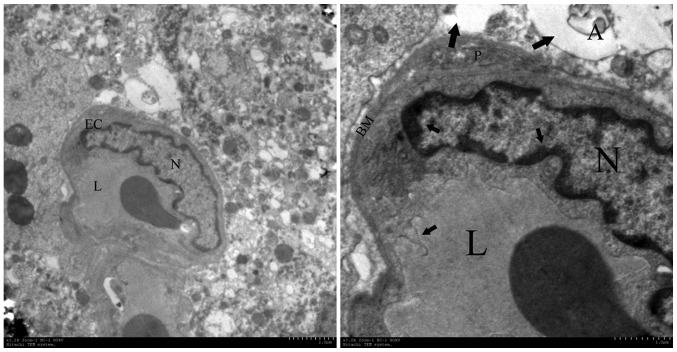
Figure 6.
Ultrastructural observations of brain capillary endothelial cells. The most pronounced alterations were observed in disturbed vessels and perivascular structures in the irradiated tissue at week 16, which contained enlargement of endothelial nuclei, pykno-chromatin (short arrow), thickening of the capillary basement membrane, folding of the plasma membrane (short arrow) and astroglial edema (long arrow). A, astrocyte; EC, endothelial cells; N, nuclei, L, capillary lumen; BM, basement membrane. Magnification: Left, ×10,000; right, ×50,000.

