Figure 7.
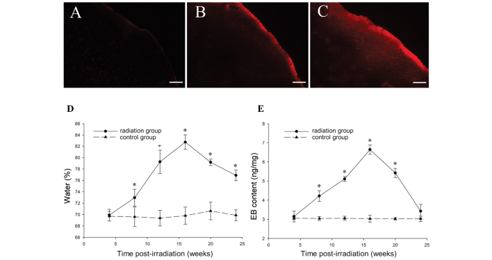
Representative results of brain water content and EB extravasation performed between 4 and 24 weeks after GKS. EB extravasation in the cortex area ipsilateral to radiation was identified by fluorescence microscopy (A) 4, (B) 8 and (C) 16 weeks after GKS. The dynamic changes in the (D) brain water content and (E) EB extravasation over time in irradiated tissues or controls are shown. Scale bar=100 μm. The data are presented as the mean ± standard deviation. *P<0.05, compared with the sham group. EB, Evans Blue; GKS, Gamma knife surgery.

