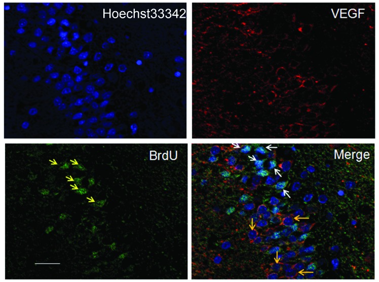
Figure 5.
Immunostaining images of BrdU- and VEGF-positive cells seven days following transplantation of BrdU-labeled hBMSCs into the cortex of rat brains (bar, 20 μm). The cell nuclei are dark blue. VEGF is stained red and was expressed around the karyotheca or in the cytoplasm. The hBMSCs (yellow arrows in the non-merged image, white arrows in the merged image) were smaller than the neurocytes, with elliptical or round nuclei that were stained green due to BrdU labeling. Orange arrows point to VEGF-positive cells, which tended to have dark blue-stained nuclei, indicating a lack of BrdU staining and therefore, that these were original host cells. VEGF, vascular endothelial growth factor, hBMSCs, human bone marrow stromal cells. BrdU, 5-bromo-2-deoxyuridine.

