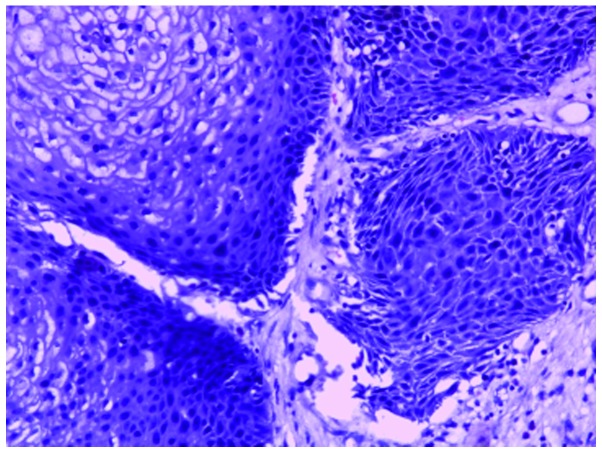
Figure 3.
Representative image of sinonasal inverted papilloma with atypical hyperplasia. The section shows the transitional cell growth of papillary epithelial cells. The cells have a disordered arrangement and the nuclei are of different sizes. Atypic epithelial cells and severe dysplasia are evident (hematoxylin and eosin staining; magnification, ×100).

