Figure 1.

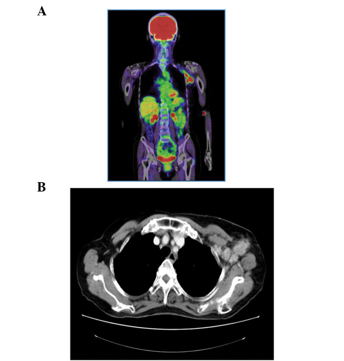
Position electron tomography (PET) and contrast-enhanced computed tomography (CT). (A) Fludeoxyglucose (FDG)-PET: FDG accumulation only in the left axillary lymph node. (B) Contrast-enhanced CT: Swelling in the left axillary lymph node.

Position electron tomography (PET) and contrast-enhanced computed tomography (CT). (A) Fludeoxyglucose (FDG)-PET: FDG accumulation only in the left axillary lymph node. (B) Contrast-enhanced CT: Swelling in the left axillary lymph node.