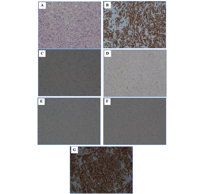
Figure 3.
Hematoxylin and eosin (H&E) staining, and various types of immunostaining of the left axillary lymph node mass. (A) H&E; (B) cytokeratin (CK)7; (C) CK20; (D) gross cystic disease fluid protein 15 (GCDFP15); (E) estrogen receptor (ER); (F) progesterone receptor (PgR); and (G) human epidermal growth factor receptor 2 (HER2). The following immunostaining results were obtained: CK7 positive (+), CK20 negative (−), GCDFP15 (partial +), ER (−), PgR (−) and HER2 score (3+). Magnification, ×100.

