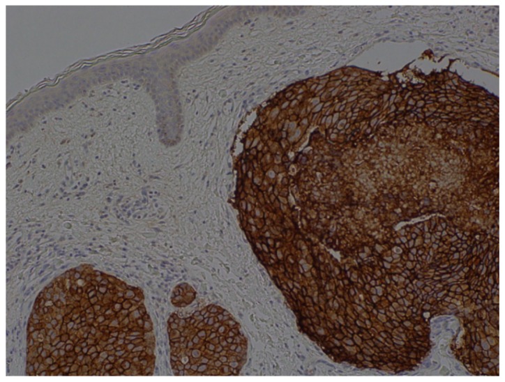
Figure 5.

Histopathological observations of a skin biopsy (human epidermal growth factor receptor 2 staining; 3+) revealed an adenocarcinoma. Atypical cells with large nuclei were detected in the blood and lymphatic vessels of the dermis and the vessels contained atypical cells (magnification, ×100).
