Figure 4.

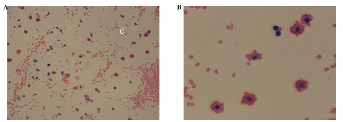
T lymphocytes (stained blue) surrounded by sheep red blood cells (stained red), forming rosettes (stained by Wright’s method), at a (A) magnification, ×400 and (B) magnification, ×800 (amplified from C).

T lymphocytes (stained blue) surrounded by sheep red blood cells (stained red), forming rosettes (stained by Wright’s method), at a (A) magnification, ×400 and (B) magnification, ×800 (amplified from C).