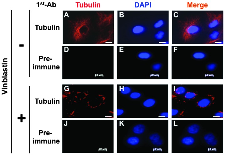
Figure 1.
Paracrystal formation in human A549 cells. A549 cells were treated with a vinblastine solution and fixed. The cells were stained with an anti-tubulin antibody followed by an appropriate secondary antibody. Nuclei were stained with DAPI. Images were acquired using an Axiovert 200M microscope. Bar = 10 μm.

