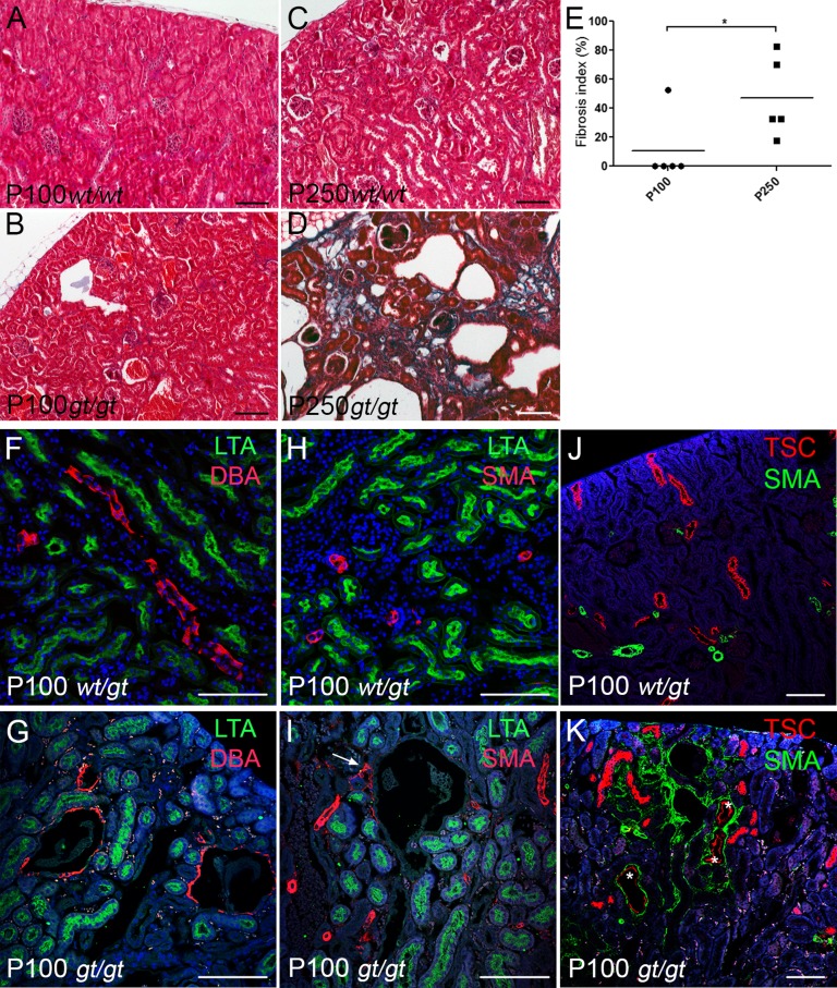
Figure 3.
Sdccag8gt/gt mice develop fibrosis. (A–D) Masson trichrome staining reveals no collagen deposits in Sdccag8gt/gt kidneys at P100 (B); however, extensive fibrosis is visible in P250 Sdccag8gt/gt kidneys (D). (E) The fibrosis index is significantly increased in Sdccag8gt/gt mice between P100 (mean 10.50±10.50) and P250 (mean 47.00±12.41; *P<0.05). (F and G) LTA-FITC and DBA-rhodamine staining on kidney sections demonstrates the formation of cysts in the CCDs (DBA) and not in the proximal tubules (LTA) of Sdccag8gt/gt kidneys at P100. (H and I) Immunofluorescence staining with α-SMA-rhodamine and LTA-FITC demonstrate the presence of myofibroblasts in the proximity of a nascent cyst (I, white arrow). (J and K) Immunofluorescence staining with αSMA-FITC and DCT marker TSC show that besides CCDs, cysts also originate from DCTs (marked with white asterisks) in P100 Sdccag8gt/gt kidneys (K). αSMA-FITC staining shows the myofibroblasts surrounding the affected tubules. wt, Sdccag8 wild-type allele; gt, Sdccag8 gene-trap allele; α-SMA, α-smooth muscle actin. Bar, 100 μm in A–D; 100 μm in F–K.

