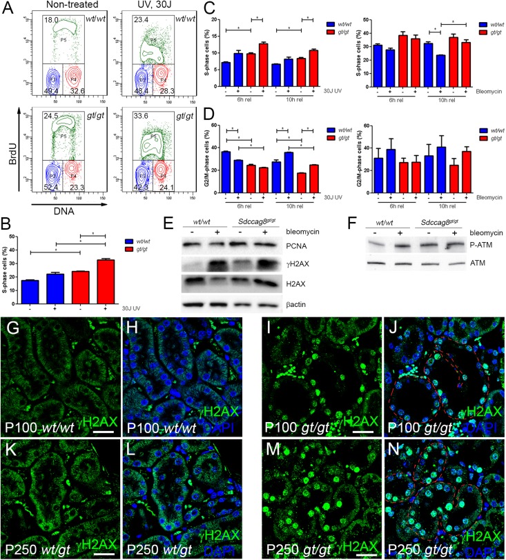
Figure 5.
Sdccag8 inactivation causes S-phase delay and replication stress. (A and B) The cell cycle profile of Immorto;Sdccag8wt/wt and Immorto;Sdccag8gt/gt nonsynchronized cells demonstrates significant accumulation of Sdccag8gt/gt cells in the S phase (P<0.05; n=3) compared with the wild-type control (P<0.05, n=3). The number of cells in the S phase is further increased after treatment with 30 J UV light. (C) Sdccag8gt/gt cells synchronized with double thymidine block show a delay in S-phase progression, which is further increased after treatment with UV light. (D) Sdccag8gt/gt cells are competent in G2/M check point activation, both in response to UV light and bleomycin treatment. (E) Sdccag8gt/gt cells have increased PCNA levels, reflecting their extended duration in the S phase. PCNA levels do not change in response to bleomycin treatment, which is in agreement with the cell cycle analysis data. Increased γH2AX levels in cultured Sdccag8gt/gt cells are a sign of replication stress. H2AX and β-actin are used as loading controls. (F) Phosphorylated ATM levels are low in cultured wild-type cells, which become elevated upon bleomycin treatment. By contrast, Sdccag8gt/gt cells display high phosphorylated ATM levels even without treatment. ATM is used as a loading control. (G–J) Immunofluorescence staining with anti-γH2AX antibody on P100 Sdccag8wt/wt and Sdccag8gt/gt kidneys shows no γH2AX staining in control kidneys (G and H), whereas the dilated tubules (red dashed lines) in Sdccag8gt/gt kidneys are positive for γH2AX (I and J). (K–N) P250 Sdccag8wt/gt kidneys are negative for gH2AX staining (K and L), while Sdccag8gt/gt kidneys show positive staining in dilated and nondilated nephron epithelium (red dashed lines) and fibrotic mesenchyme (M and N). wt, Sdccag8 wild-type allele; gt, Sdccag8 gene-trap allele; PCNA, proliferating cell nuclear antigen. Bar, 25 μm in G–J; 20 μm in K–N.

