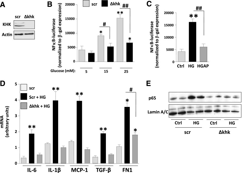
Figure 6.
Fructose metabolism blockade is associated with reduced inflammation in proximal tubular cells exposed to high glucose levels. (A) Representative Western blot of HK-2 cells control (Scr [scramble]) and stably silenced for KHK expression (Δkhk). (B) Luciferase units—normalized to β-gal expression—in scr and Δkhk cells transfected with an NF-κB-luciferase reporter cassette and exposed to increasing levels of glucose. (C) Luciferase units—normalized to β-gal expression—in regular HK-2 cells transfected with an NF-κB-luciferase reporter cassette and exposed to high glucose (HG; 25 mM) in the presence or absence of allopurinol (100 µM). (D) Quantification of mRNA levels of cytokines (IL-1β and IL-6), chemokines (MCP-1), and fibrotic genes (TGFβ and FN1) in scr and Δkhk HK-2 cells exposed to high glucose levels (25 mM). (E) Representative Western blot of p65 from nuclear extracts from scr and Δkhk cells under control (ctrl) or high glucose conditions. Mean±SEM. *P<0.05 and **P<0.01 versus respective control; #P<0.05 and ##P<0.01.

