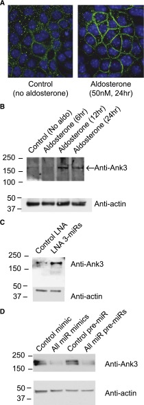
Figure 6.
Ank3 is regulated by aldosterone and miRs. (A) Immunofluorescent images of mCCD cells cultured on filter supports and labeled for Ank3 (green) in cells without (left) and following aldosterone stimulation (right). Nuclei are counterstained in blue (DAPI); white bars=10 μm. (B) Western blots of Ank3 expression from whole cell lysates of mCCD cells cultured on filters and stimulated with aldosterone for 6, 12, and 24 hours. (C) Expression of the three miRs was reduced by LNA transfection (as in Figure 4) and cells seeded onto filters. A Western blot of Ank3 expression from whole cell lysates in control (unstimulated) and LNA transfected, miR reduced mCCD cells (no aldosterone) is presented. (D) The three miRs were overexpressed using miR mimics or plasmid pre-miR transfection. Expression of Ank3 from whole cell lysates is presented in the Western blot following miR overexpression or in control transfected mCCD cells. In both cases, miR overexpression reduced endogenous Ank3 expression.

