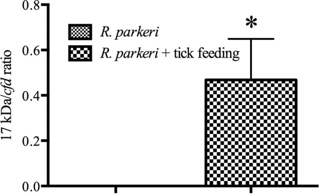
Fig. 1.
qPCR for R. parkeri 17 kDa antigen gene relative to mouse cfd in the skin at 8 dpi. Relative quantification was used to account for variation in weight of tissues at the time of nucleic acid extraction. The mean R. parkeri numbers ± SEM as detected in mice that either (A) had no nymphal A. maculatum infestation or (B) had nymphal A. maculatum feeding at the inoculation site (* denotes significance between groups of P ≤ 0.05).

