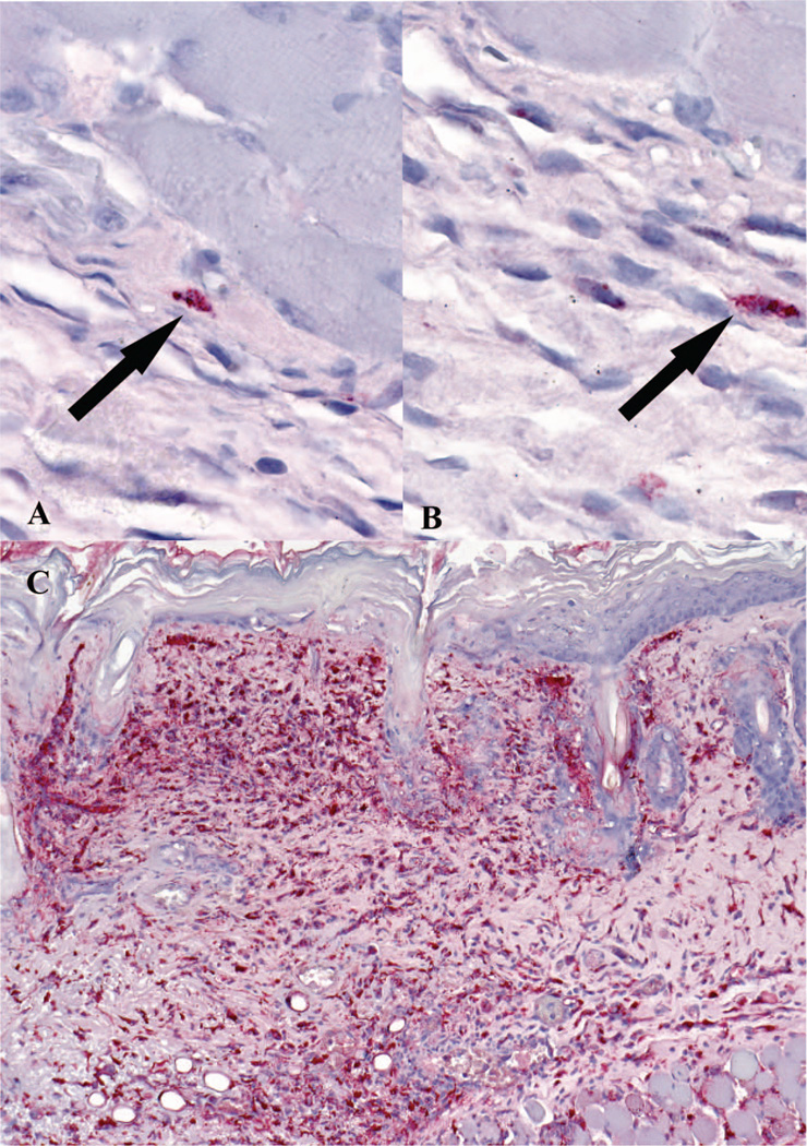
Fig. 2.
Immunohistochemical detection of SFG Rickettsia at tick feeding sites. A and B represent cutaneous tissues of the buffer-inoculated mice with tick feeding group. Arrows indicate rare positive cells (red) for SFG Rickettsia. Frame C displays florid staining of SFG Rickettsia in the cytoplasm of histiocytic cells and endothelial cells in the skin of R. parkeri inoculated mice with tick feeding at 8 dpi. Immunoalkaline phosphatase technique with naphthol-fast red and hematoxylin counterstain.

