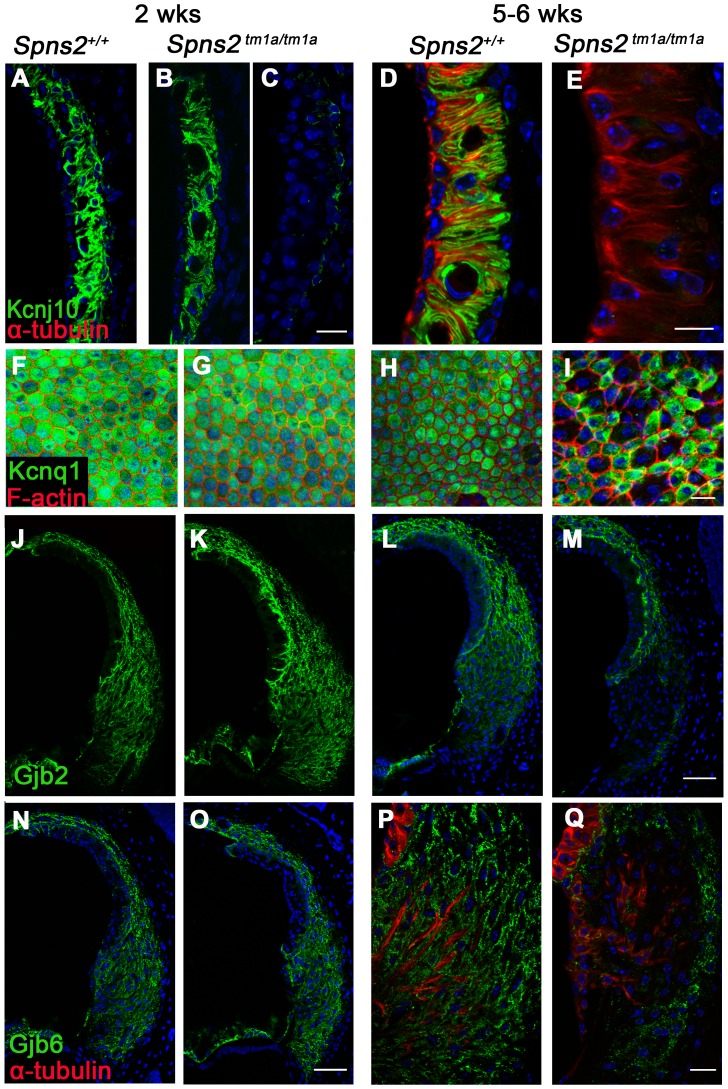
Figure 8. Progressive decrease in expression of Kcnj10, Kcnq1, Gjb2 and Gjb6 in Spns2tm1a/tm1a mice.
A–E: At P14, Kcnj10 expression (green) of homozygotes was comparable to that of wildtype in apical turns, while in basal turns, some appeared normal (B) as wildtype (A), but some appeared largely reduced (C). At 5–6 weeks old, Kcnj10 labelling was absent in homozygotes (E). Acetylated α-tubulin (red) was used to label strial marginal cells in D,E. F–I: Whole mount preparations of the stria. Kcnq1 labelling (green) was detected at P14 in both homozygotes (G) and wild types (F), but it was absent from those marginal cells with enlarged cell boundaries in Spns2tm1a/tm1a mice at 5–6 wks (I). Phalloidin (red) labelled filamentous actin to reveal the boundaries of marginal cells. J–Q: Gjb2 and Gjb6 were present in the fibrocytes of the spiral ligament in both wild type and Spns2tm1a/tm1a mice at P14 (J,K,N,O). At 5–6 wks, expression was absent in the area behind the spiral prominence corresponding to the type II fibrocytes in homozygotes (M and Q) compared with wildtypes (L and P) of the same age. Root cells were labelled by acetylated α-tubulin (red) in P,Q. DAPI (blue) labelled the nuclei. Scale bar, 10 µm in D,E. 20 µm in A–C, F–I,P,Q. 50 µm in J–O.

