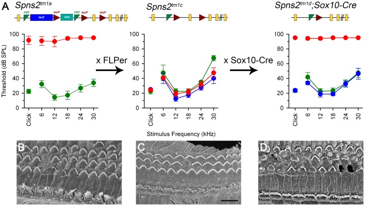
Figure 9. ABR thresholds and SEM assessment suggest a local function of Spns2 in the inner ear.
ABR thresholds (means +/−SD) are shown for homozygotes (red), heterozygotes (blue) and wildtypes (green), aged 7–14 weeks. Mice homozygous for the Spns2tm1a allele displayed elevated ABR thresholds and degeneration of hair cells (A left, 4 wks and B). By crossing with mice expressing Flp recombinase to excise the inserted cassette, Spns2tm1c/tm1c mice were produced, which had normal ABR thresholds and normal hair cell morphology (A middle, 8 wks and C). Then Spns2tm1c/tm1c were crossed with Sox10-Cre mice to produce Spns2tm1d/tm1d;Sox10-Cre mice which showed no response up to 95 dB SPL and hair cell degeneration with bulges and holes in the reticular lamina (A right, 4 wks and D). SEM images are taken from the middle turn (40–70%) of the cochlea. Scale bar: 10 µm in B,C,D.

