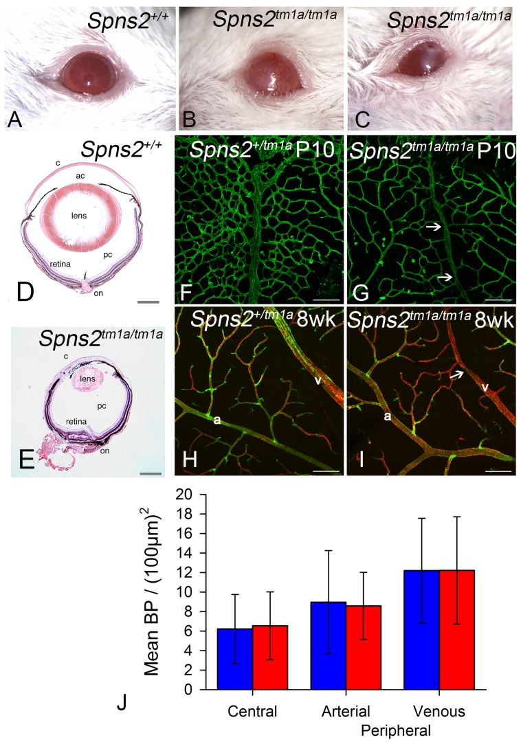
Figure 12. Eye defects in Spns2tm1a/tm1a mice.
A–C, Phenotype screening was performed on 15 week old Spns2tm1a/tm1a mice and identified buphthalmos, corneal opacity with vascularization and possible ulceration (B) and corneal opacity with vascularisation and polyp, thick discharge, elongated pupil which did not fully dilate with tropicamide (C). Albino mice were selected to achieve clearer demonstration, and a wildtype is shown in (A). D, Pupil-optic nerve section of a 15 week old wildtype eye. E, The Spns2tm1a/tm1a mice showed a grossly abnormal eye with corneal opacity, vascularization, collapsed anterior chamber, small cataractous lens, and focal retinal degeneration. c = cornea, ac = anterior chamber, pc = posterior chamber, on = optic nerve. Scale bar = 450 µm. F,G,H,I Analysis of retinal vasculature was performed on retinal wholemounts from P10 pups (F,G) and 8 week old adult mice(H,I). Retinal vasculature was stained with Isolectin B4 (green) to visualise the endothelium and Proteoglycan NG2 (red) to visualise pericytes. Whereas arteries (a) appeared morphologically normal, veins (v) appeared thinner in Spns2tm1a/tm1a (I) than in Spns2+/tm1a (H) and had an irregular caliber with regions of narrowing (arrows). Although the retina has three capillary plexi only the primary plexus is shown at both P10 and 8 weeks as this is the first plexus to form and mature. Scalebars: 50 µm. J, Branch point analysis was performed on P10 retinal vasculature (F,G) to determine whether retinal vasculature showed any developmental abnormalities in vascular patterning. No significant difference was detected between Spns2tm1a/tm1a (red) and Spns2+/tm1a (blue) mice in the central retina (mature vessels) or the periphery, where the vessels are still developing at P10, in either the arteries or veins (Mann-Whitney U test for central branch points, p = 0.69; t-test for peripheral branch points, arterial p = 0.899, venous p = 0.996).

