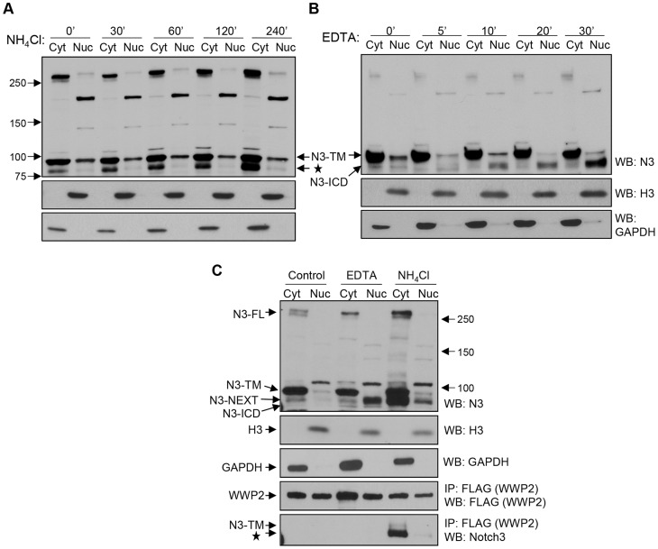
Figure 4. WWP2 interacts with endogenous Notch3 in cancer cells.
(A) MCF7 cells were incubated with 25 mM NH4Cl for 0, 30, 60, 120, and 240 minutes. Cells were lysed and the lysates separated into nuclear (N) and cytosol/membrane (C) fractions. Western blot analysis was performed with an anti-Notch3 antibody. Equal loading was determined with an anti-GAPDH antibody for the cytosol/membrane fraction and with an anti-histone 3 antibody for the nuclear fraction. The star represents the N3-NEXT bands. (B) MCF7 cells were incubated with 2.5 mM EDTA for different times as indicated to induce Notch-ICD generation. Cells were lysed and fractionated into nuclear (N) and cytosol/membrane (C) fractions. Western blot analysis was performed with an anti-Notch3 antibody. (C) Ovarian cancer cells were first transfected with the flag tagged WWP2 expressing plasmids. The cells were then treated with 2.5 mM EDTA for 20 minutes or with 25 mM NH4Cl for 240 minutes, and then fractionated into nuclear and cytosol/membrane fractions. Immunoprecipitation (IP) was performed with anti-flag agarose beads for pull-down and a rabbit anti-Notch3 antibody for western blot. Expression of GAPDH and histones were used for demonstrating the purity of nuclear and cytosol/membrane fractions, respectively.

