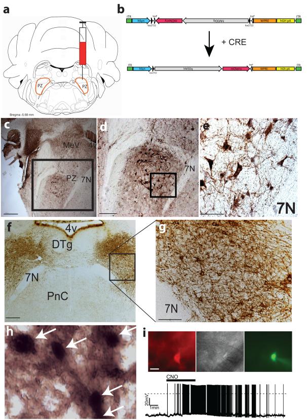
Figure 1. Cre–dependent expression of the hM3Dq receptor in PZ GABAergic neurons.
(a) coronal section outline shows the injection target (delimited node of PZ GABAergic neurons) in Vgat–IRES–cre mice. (b) details of hSyn–DIO–hM3Dq–mCherry–AAV (hM3Dq–AAV) vector injected. (c) GFP immunolabeling in the brain of Vgat–IRES–cre, lox–GFP mice shows the location of GABAergic (VGAT+) PZ neurons (scale bar = 500 μm); (d) higher power photomicrograph of GABAergic PZ neurons targeted for injection (scale bar = 250 μm); (e) morphology of magnocellular PZ GABAergic neurons (scale bar = 65 μm); (f) bilateral expression (brown immunoreactivity in neuropil) of the hM3Dq receptor in PZ GABAergic neurons following AAV–mediated transduction (scale bar = 300 μm). (g) expression of hM3Dq receptors is evident on the cell surface and processes of GABAergic PZ soma (scale bar = 70 μm). (h) high magnification image showing red–brown cytoplasmic and neuropil immunostaining with black nuclear c–Fos immunoreactivity indicates excitation of GABAergic hM3Dq+ PZ neurons by CNO (scale bar = 20 μm). (i) CNO (500 nM bath applied) produced depolarization and firing in hM3Dq–expressing GABAergic PZ neurons in brain slices. 4v: fourth ventricle; 7n: facial nerve; Cre: cre–recombinase; CNO: clozapine–N–oxide; DTg: dorsal tegmental nucleus; PnC: pontine reticular nucleus; PZ: parafacial zone.

