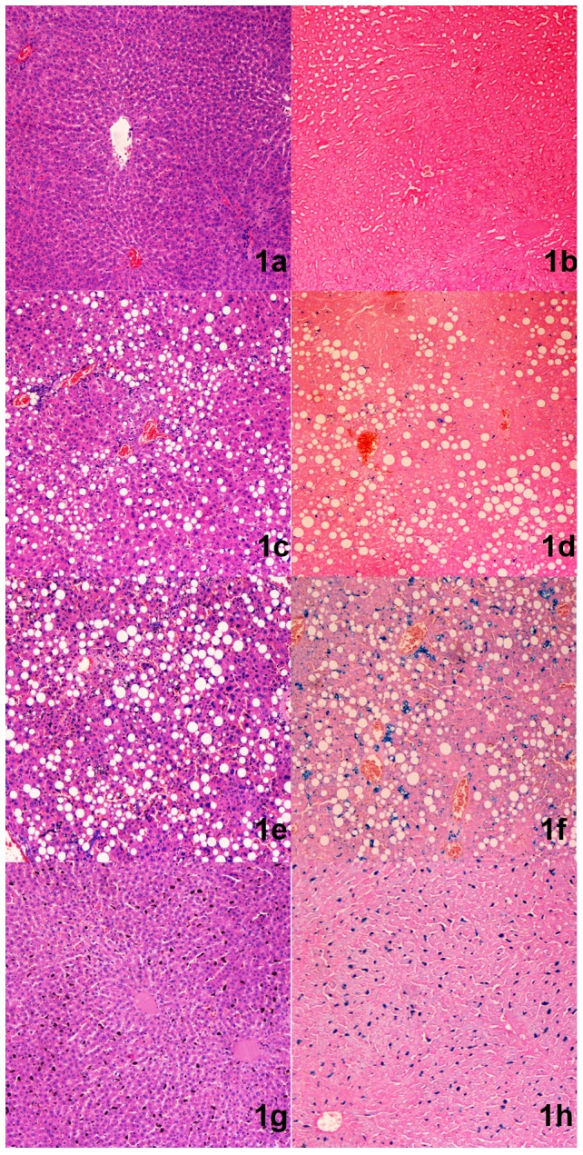
Figure 1. The rats were grouped by their liver histological results.

Normal group: 1a) No lipid droplets in hepatocytes in the field of vision with HE staining. 1b) Prussian blue staining; there was no blue stained material in the field of vision, indicating the absence of iron deposition. Fatty liver group: 1c) Many hepatocytes were affected by lipid droplets on HE staining. 1d) Prussian blue staining resulted in no blue stained material, indicating the absence of iron deposition. Coexisting group: 1e) Many hepatocytes were affected by lipid droplets with HE staining. 1f) There were blue stained dots with Prussian blue staining, indicating significant iron deposition in this rat. Liver iron group: 1g) No hepatocytes were affected by lipid droplets on HE staining. 1h) There are blue stained dots in Prussian blue staining, indicating significant iron deposition in this rat.
