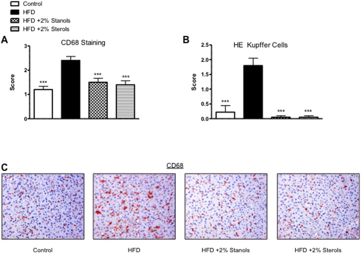
Figure 3. Foamy Kupffer Cells.
(A) Scoring for the size and foamy appearance of Kupffer cells using CD68 staining. (B) Scoring for the size and foamy appearance of Kupffer cells using HE staining. A score ranging from 0–3 was given by an experienced pathologist. (C) Representative pictures of the foamy Kupffer cell appearance with CD68 staining (200x magnification). *P<0.05, **P<0.01, and ***P<0.001, respectively.

