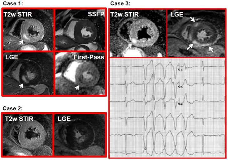
Figure 1. Examples of HCM patients.
Case 1: a patient with HCM presenting with HyT2 (arrow in T2-STIR image), myocardial fibrosis (arrow LGE image), and pefusion defect (arrow in the frame of the first pass gadolinium) in the same myocardial segments; Case 2: a patient with HCM having myocardial fibrosis (LGE image) without HyT2 (T2-STIR image); Case 3: a patient with HyT2 (arrow in T2-STIR images) and myocardial fibrosis (arrow in LGE images) having a run of NSVT in the ECG stripe (lower panel).

