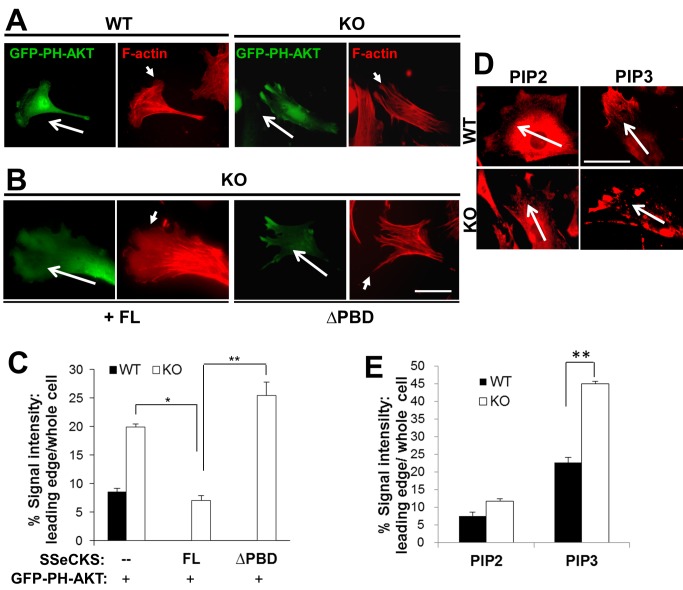
Figure 4. SSeCKS attenuates PIP3 enrichment at the leading edge.
A, Chemotactic WT or KO MEF (long arrows: chemotaxis direction) transiently expressing the PIP3 reporter, GFP-PH-AKT, and stained for F-actin, showing enrichment of PIP3 in the leading edge filopodia of KO cells. Short arrows, leading edge lamellipodia (WT cells) or filopodia (KO cells). B, Re-expression of FL-, but not ΔPBD-, SSeCKS-GFP rescues leading edge lamellipodia formation in KO MEF and suppresses leading edge enrichment of the GFP-PH-AKT reporter. Scale bar, 10 µm. C, Quantification of leading edge GFP-PH-AKT levels (normalized to total cell GFP) determined for 3 fields containing at least 10 cells/field in 2 independent experiments. Error bars, S.E. *, p<0.01, **, p<0.005. D, PIP2 or PIP3 in chemotactic leading edges of WT or KO MEF by IFA or quantified as in E. **, p<0.01. Scale bar, 10 µm.

