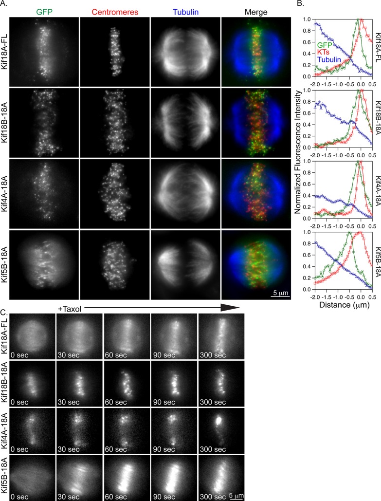
FIGURE 2:
Kif18A-tail chimeras rapidly accumulate at K-fiber ends in Taxol-treated cells. (A) Fluorescence micrographs of the indicated GFP-tagged kinesins (green) in Kif18A-depleted HeLa cells after a 5-min incubation with 10 μM Taxol. Cells were immunostained with ACA (centromeres, red) and tubulin (blue). (B) Line scans of GFP-tagged kinesins (green trace) relative to tubulin (blue trace) and ACA (red trace) along K-fibers in 10 μM Taxol–treated cells. (C) Still images from live-cell analyses of GFP-tagged kinesin relocalization after the addition of Taxol. Taxol was added immediately after the 0-s image was captured.

