FIGURE 6:
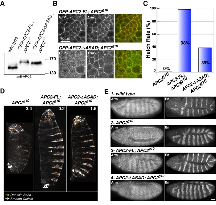
APC2 self-association is required to negatively regulate Wnt signaling in the Drosophila embryo. (A) Immunoblot of 0- to 6-h embryonic lysates demonstrates that the level of expression of GFP-APC2-FL and GFP-APC2-ΔASAD is comparable to that of endogenous APC2. (B) GFP-APC2-FL is enriched at the cell cortex with Arm in embryonic epithelia, whereas GFP-APC2-ΔASAD is primarily cytoplasmic. Scale bar, 10 μm. (C, D) Expression of GFP-APC2-FL rescued the lethality of APC2-null (APC2g10) embryos and restored the wild- type cuticle phenotype, whereas the APC2-ΔASAD mutant only moderately rescued the lethality and cuticle phenotype. The numbers in D indicate the phenotypic average for each genotype (scoring criteria as in McCartney et al., 2006). Cuticle images are shown at the same scale. (E) Representative embryos showing Arm and En protein expression in wild-type (1) and APC2-null (2) embryos. APC2-FL restored wild-type Arm levels and the En expression domain of APC2-null (APC2g10) embryos. APC2-ΔASAD weakly suppressed Arm accumulation and restored a weak Arm stripe pattern in the epidermis. The En expression domain remains expanded in APC2-null embryos expressing APC2-ΔASAD. Scale bar, 25 μm.

