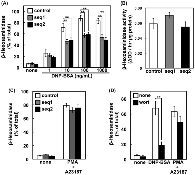
Figure 2. β-hexosaminidase release of PI3K-C2α-knockdown cells.
(A) FcεRI-mediated β-hexosaminidase release. Control or PI3K-C2α-knockdown cells were sensitized with anti-DNP IgE and then stimulated with the indicated concentrations of DNP-BSA at 37°C for 10 min. After centrifugation, the β-hexosaminidase activity in the supernatant was determined and is shown as a percentage of the activity in the total cell lysate. The data are shown as the means ± s.d. (n = 4). (B) Content of β-hexosaminidase. The cells were solubilized for determination of β-hexosaminidase activity. The activities are expressed as the absorbance change at 405 nm per 1 µg of protein and are shown as the means ± s.d. (n = 4). (C) PMA/A23187-induced β-hexosaminidase release. The cells were pre-treated with 30 nM PMA at 37°C for 10 min and then stimulated with 1 µM A23187 for 10 min. The β-hexosaminidase activity was measured as in (A). The data are shown as the means ± s.d. (n = 4). (D) Effect of wortmannin. Sensitized or non-sensitized RBL-2H3 cells were incubated at 37°C with or without 30 nM PMA for 10 min. When added, 1 µM wortmannin was included during this period. The cells were then incubated with or without 100 ng/mL DNP-BSA or 1 µM A23187 for 10 min. The β-hexosaminidase activity was measured as in (A). The data are shown as the means ± s.d. (n = 3).

