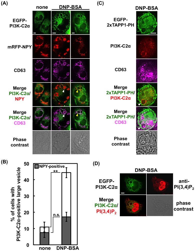
Figure 6. Existence of PI3K-C2α and PtdIns(3,4)P2 on large vesicles in FcεRI-stimulated RBL-2H3 cells.
(A) Existence of PI3K-C2α on CD63-positive large vesicles containing or not containing NPY-mRFP. RBL-2H3 cells were transfected with EGFP-PI3K-C2α and NPY-mRFP. The cells were fixed before or after FcεRI stimulation, and stained with anti-CD63 and Alexa 647-labeled anti-mouse IgG antibodies. The arrowheads indicate the EGFP-PI3K-C2α and CD63-positive vesicles; the white indicates the NPY-mRFP-containing vesicle, while the yellow indicates the empty vesicle. Scale bar = 5 µm. (B) Numbers of cells containing EGFP-PI3K-C2α and CD63-positive large vesicles (>2.5-µm diameter). Solid bar indicates the number of cells possessing one or more vesicles containing NPY-mRFP. For each experimental condition, 106 cells were analyzed. The data are shown as the means ± s.e.m. from three separate experiments. (C) Existence of endogenous PI3K-C2α and PtdIns(3,4)P2 on CD63-positive large vesicles. RBL-2H3 cells were transfected with EGFP-2×TAPP1-PH. After stimulation, the cells were fixed, treated with anti-PI3K-C2α and anti-CD63, and then stained with Alexa 555-labeled anti-rabbit and Alexa 647-labeled anti-mouse IgG antibodies. The arrowhead indicates the EGFP-2×TAPP1-PH, PI3K-C2α and CD63-positive vesicle. Scale bar = 5 µm. (D) Existence of PI3K-C2α and PtdIns(3,4)P2 on large vesicles. RBL-2H3 cells were transfected with EGFP-PI3K-C2α. After stimulation, the cells were fixed and stained with anti-PtdIns(3,4)P2 and Alexa 647-labeled anti-mouse IgG antibodies. Scale bar = 5 µm.

