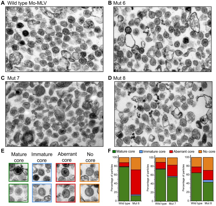
Figure 4. Analysis of the Mo-MLV p12 mutant intra-virion CA core by transmission electron microscopy.
Purified Mo-MLV VLPs were pelleted and prepared for TEM. (A–D) Representative electron micrographs of wild type Mo-MLV and p12 mutant VLPs are shown. (E) Representative examples of the different core morphologies used to classify particles observed in the micrographs. (F) The morphology of all Mo-MLV particles (within 80–120 nm in diameter) were scored from multiple micrographs (at least 93 particles scored for each) and the results are displayed as a percentage of the total particles scored. All scale bars are 50 nm.

