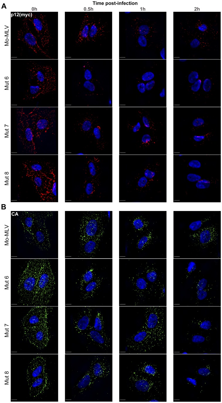
Figure 6. Immunofluorescence of p12 and CA in cells infected with Mo-MLV p12 mutants.
U/R cells were challenged with ecotropic wild type or p12 mutant Mo-MLV VLPs, containing a myc-tag in p12, by cold spinoculation (MOI 3). Cells were fixed at various times post-infection and stained with either an (A) anti-myc or (B) anti-CA antibody followed by a Cy3 (A) or FITC (B) -conjugated secondary antibody. The nuclear DNA was counterstained using DAPI (blue). Images from the time course were captured using a spinning disk confocal microscope and representative images of cells from each time point are shown. All images are three dimensional acquisitions projected on a two dimensional plane. Images were processed using SlideBook. Scale bars are 10 µm.

