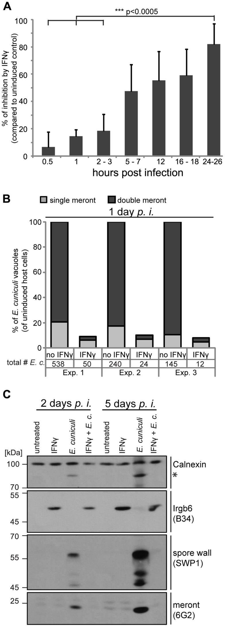
Figure 1. IFNγ restricts E. cuniculi growth in mouse embryonic fibroblasts.
(A) Mouse embryonic fibroblasts (MEFs) from C57BL/6 mice were induced with IFNγ for 24 h or left uninduced before infection with E. cuniculi spores. Cells were fixed at the indicated time points and the number of meronts (stained with anti-meront mAb 6G2) per 500 host nuclei (stained with DAPI) was counted. The inhibition in the IFNγ-treated sample compared to the uninduced control sample is presented as mean +/− standard deviation (SD) of 3–7 replicates per time point from at least 2 individual experiments. Significant differences (of 0.5 h, 1 h and 2–3 h compared to 24–26 h) were calculated with a two tailed T-test. (B) MEFs were induced with IFNγ or left uninduced, infected with E. cuniculi spores for 24 h and stained as in A. Single meronts and meronts that divided once (double meront) were counted per 500 host nuclei and shown as percent of total vacuoles of uninduced controls. Numbers indicate the counted number of single or double meronts per 500 host cells. Data from three independent experiments (Exp. 1–3) is presented. (C) MEFs were stimulated with IFNγ and/or infected with E. cuniculi spores for 2 or 5 days or left untreated. Cell lysates were separated by SDS-PAGE and Western Blots were cut into three regions to probe for anti-meront mAB 6G2 as well as anti-spore wall protein 1 pAS SWP1. Calnexin staining served as loading control and Irgb6 staining (mAB B34) to proof IFNγ-induction. The asterisk marks an unknown E. cuniculi-derived protein that is detected by the Calnexin antibody. These Western Blots emerged from one single SDS-PAGE, the 45–70 kDa region was first probed with mouse mAB B34, stripped, and then probed for anti-SWP1 rabbit pAS. Experiments for both time points were performed at least three times.

