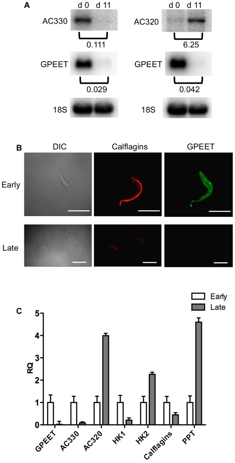
Figure 6. Differential expression of markers in early and late procyclic culture forms.
(A) Northern blot analysis of adenylate cyclases Tb927.5.330 (AC 330) and Tb.927.5.320/285b (AC 320). GPEET was used to verify that the cultures correspond to early and late procyclic forms, respectively. Signals were quantified using a PhosphoImager and normalised against 18S rRNA as described previously [47]. Signals in early procyclic forms were set at 1. (B) Immunofluorescence analysis reveals that GPEET and calflagin are co-expressed by early procyclic culture forms and are not detectable in late procyclic culture forms. Scale bar: 10 µm. (C) Quantitative RT-PCR performed using RNA from early procyclic culture forms and late procyclic culture forms 11 days after removal of glycerol from the medium [4], [7]. RQ: Relative quantification. Expression levels in early procyclic forms are set at 1. α-tubulin was used to normalise mRNA levels. Error bars are ΔCt standard errors. AC 330: Tb927.5.330 3′ UTR; AC 320: Tb927.5.320/285b 3′ UTR; HK1: 3′ UTR of Tb927.10.2010: HK2: 3′ UTR of Tb927.10.2020; Calflagin: coding region of Tb927.8.5460, Tb927.8.5440, Tb927.8.5465, Tb927.8.5470. PPT: coding region of Tb927.1.2850, Tb927.1.2880 (putative pteridine transporters).

