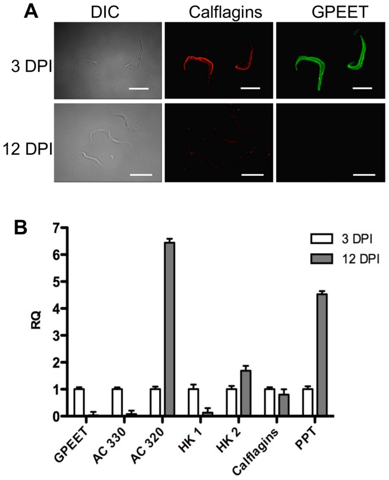
Figure 7. Differential expression of early and late procyclic form markers in tsetse flies.
A. GPEET and calflagin are co-expressed by early procyclic forms 3 days post infection (DPI), but neither is detectable in late procyclic forms 12 DPI. Trypanosomes were isolated from tsetse fly midguts, fixed with formaldehyde and glutaraldehyde and permeabilised with Triton-×100. Immunofluorescence was performed with anti-GPEET and anti-calflagin antisera. Scale bar: 10 µm. B. Quantitative RT-PCR was performed with RNA isolated from infected tsetse flies 3 and 12 days post infection. Gene designations are the same as for Figure 6. RQ: relative quantification. α-tubulin was used to normalise mRNA levels. Error bars are ΔCt standard errors.

