Figure 1.
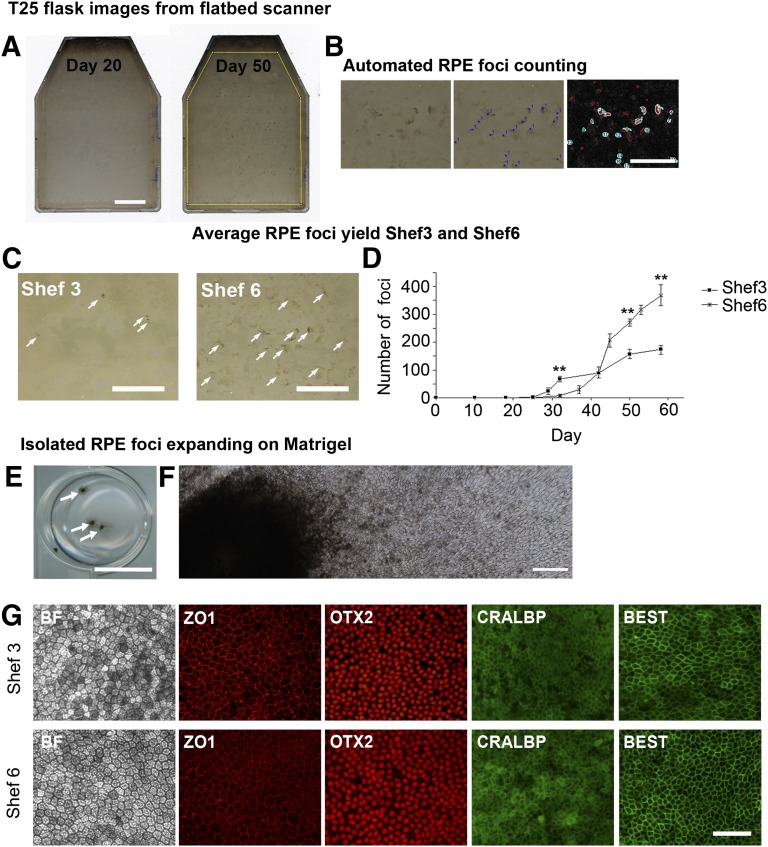
Pigmented foci of RPE begin to appear between days 25 and 32 after human embryonic stem cell seeding. (A): Images of T25 flasks acquired on a flatbed scanner showing the emergence of pigmented foci at day 50. Scale bar = 10 mm. (B): Pigmented areas are manually counted to determine the number of foci (center), with automated highlighting and counting of pigmented areas using a size and pigment intensity threshold (right). Scale bar = 5 mm. (C): Pigmented foci in typical flasks of Shef3 and Shef6 at day 50. Consistently more RPE per cm2 were present in Shef6. Scale bar = 10 mm. (D): The accumulation of individual RPE foci over time in T25 flasks of Shef3 (n = 3) and Shef6 (n = 3) was measured using a flatbed scanner and automated foci counting in ImageJ (NIH). Error bars = SEM of biological replicates. A significant difference was seen in total foci numbers over time and between the two cell lines (p < .001). (E): Manually isolated RPE expanding on a Matrigel-coated 12-well plate. Arrows highlight individual foci. Scale bar = 10 mm. (F): Stitched photomicrographs at 20× magnification depicting RPE cells expanding on Matrigel from a single, manually isolated foci. Scale bar = 500 µm. (G): Immunostaining of pigmented cells derived from Shef3 (upper panel) and Shef6 (lower panel). RPE have typical cobblestone morphology and express RPE markers ZO1, OTX2, CRALBP, and BEST. Scale bar = 100 µm. Abbreviations: BEST, bestrophin; RPE, retinal pigment epithelium.

