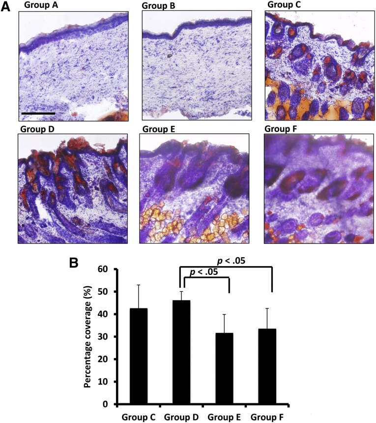
Figure 5.
Groups C, D, and E regenerated sebaceous glands, an important component of skin. (A): Oil Red O staining showed that groups A and B did not form sebaceous glands in the healed area of skin defects. Groups C, D, and E regenerated sebaceous glands, and sebum secretion was observed on the skin surface. The skin samples from nude mice (group F) also showed sebaceous glands. (B): When both microfluidic-enriched CD34-positive (CD34+) stem cells and unenriched epidermal cells were transplanted into skin defects together with dermal fibroblasts, group D had higher sebaceous gland coverage relative to group E (microfluidic-enriched CD34+ stem cells and dermal cells only) and group F (the natural skin of nude mice) (n = 10 per group, mean ± SD; p < .01, respectively). Scale bar = 250 μm (groups A–F).

