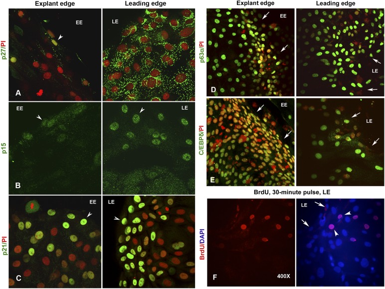
Figure 3.
Expression of cyclin-dependent kinase inhibitors in limbal explant cultures. (A): Nuclear expression of p27 (green) in few cells at the EE (left). Note the complete absence of fluorescence signal in the surrounding epithelial cells marked by PI (red). However, the epithelial cells at the LE show intense cytosolic signal for p27 (right). (B): Nuclear expression of p15 (green) only in few cells at the EE (left) and at the LE (right). Only background cytosolic fluorescence was noted in the remaining epithelial cells of the middle zone. (C): Bright nuclear expression of p21 (green) in cells localized to both the EE (left) and LE (right) cells. Expression of stem cell markers in limbal explant cultures. Epithelial cells expressing p63α (D) and C/EBPδ (E) at the EE (left) and LE (right) of explant cultures. The cells were counterstained with PI to label the nuclei in red. (F): Proliferating epithelial cells that incorporated the BrdU label (red) in a 30-minute pulse. Note the localization of BrdU-positive cells at approximately two or three cells behind the migrating or leading edge. The cells were counterstained either with PI (red) or DAPI (blue) to label the nuclei. Arrows indicate the EE and LE boundaries. Arrowheads mark the cells expressing different antigens. Magnification, ×400 for all images. Abbreviations: BrdU, 5-bromo-2′-deoxyuridine; DAPI, 4′,6-diamidino-2-phenylindole; EE, explant edge; LE, leading edge; PI, propidium iodide.

