
Fig. 3.
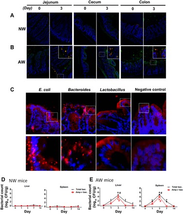
Bacterial penetration into gut mucosa and extraintestinal organs of AW mice after infection with Amp-r E. coli. A and B: jejunal, cecal, and colonic segments of NW and AW mice were subjected to fluorescence in situ hybridization with probes targeted to universal bacteria (green) and E. coli (red). Cell nuclei (blue) have been superimposed on the images. Representative photomicrographs (magnification ×200) were obtained from at least 5 mice per group. Inset: higher magnification of bacteria. C: penetration of E. coli, Bacteroides, and Lactobacillus to intestinal mucosa of AW mice on PI day 3. D and E: extraintestinal bacterial counts in liver and spleen homogenates of NW and AW mice. *P < 0.05 vs. NW; #P < 0.05 vs. day 0. n = 5/group. CFU, colony-forming units.