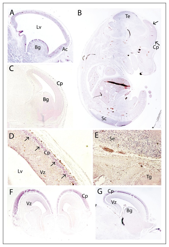
Fig. 2.
Expression pattern of miR-185 in the developing mouse brain at E12.5 (A, sagittal), E14.5 (B, D, E, sagittal), E15.5 (F, coronal) and E16.5 (G, sagittal). A negative control with a sense probe is also included (C, E14.5, sagittal). At E12.5, miR-185 is present in the developing cerebral cortex, being most pronounced in the anterior cortex (A); miR-185 is also present in the basal ganglia (A). At E14.5, miR-185 expression is prominent in the cortical plate (B, D) as well as in the tectum and the spinal cord (B). The septum also shows enriched miR-185 expression (E), with no expression in the trigeminal ganglion (E). At E15.5 (F) and E16.5 (G), miR-185 is clearly observed in the cortical plate, with low expression in the ventricular zone. Arrows indicate miR-185 expression in the mantle layer. Ac = anterior cortex; Bg = basal ganglia; Cp = cortical plate; Lv = lateral ventricle; Sc = spinal cord; Se = septum; Te = tectum; Tg = trigeminal ganglion; Vz = ventricular zone.

