
Figure 4.
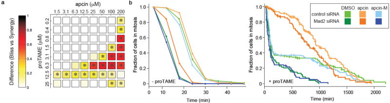
Apcin synergizes with proTAME to prolong mitotic duration. a, RPE1 cells were treated with indicated concentrations of apcin and proTAME for 18 hours, fixed, and mitotic index determined by automated high-throughput imaging. The panel shows the difference between the mitotic indices calculated by a Bliss-independence model compared to a synergy model; any positive value indicates synergy. * P<0.05 based on analysis of four technical replicates. b, Asynchronous RPE1 cells expressing H2B-GFP were transfected with siRNA and twenty-four hours later treated with apcin or apcin-M (25 μM) and/or proTAME (6 μM). Cells were then imaged every 6 minutes for 45 hours. Mitotic duration and cell fate (Extended Data Fig. 8b) were determined by manual inspection of the videos and plotted as inverse cumulative frequency (-proTAME) or Kaplan-Meier curves (+proTAME). The hatch marks on the Kaplan-Meier curves indicate censored cells that did not exit mitosis or die in mitosis before the end of the movie or before they migrated out of the field of view. Graphs include the combined results of two independent experiments.