
Fig. 4.
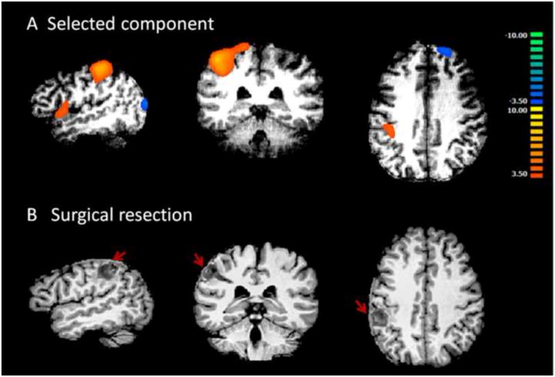
Results from patient #2. A shows the only component selected by the algorithm and accepted by visual inspection. This component has high lateralization value and is localized in the left parietal lobe. This patient had left parietal lobe epilepsy. B shows surgical resection in left parietal lobe, indicated by red arrows. The orange cluster in left parietal region shown in A agrees well with the surgical resection in B.