
Fig. 5.
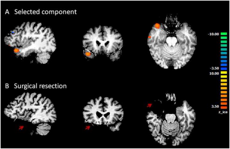
Results from Patient #3. A shows the only component selected by the algorithm and accepted by visual inspection. This component is located at the anterior portion of the left temporal lobe. This patient had left temporal lobe epilepsy. B shows surgical resection in left temporal lobe. Orange cluster in left temporal lobe in A falls well within the surgical resection indicated in B.