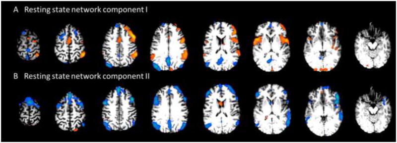
Fig 6.

Results from Patient #8. A shows one component associated with the bilateral fronto-parietal association cortex, which represents a typical resting state network. Additionally, there are also clusters on the right lateral frontal and parietal regions. B shows another component associated with the bilateral network, with an additional cluster on the right fronto-parietal region. This patient was initially diagnosed with right parietal epilepsy. Presurgical ECoG revealed diffuse seizure onset involving a large region simultaneously. This patient was not selected to receive surgical resection.
