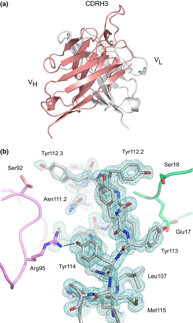
Figure 5.

(a) Overall structure of the M0418 scFv. The disposition of CDRH3 above the surface of molecule A is shown, and the H and L chain V domains are coloured in salmon and white, respectively. (b) CDRH3 from molecule A. A 2Fo-Fc electron density map is contoured at 1.0σ around residues from CDRH3. Two symmetry-related copies of molecule B (pink and green) flank CDRH3 on either side. Residues from CDRH3 molecule A are shown as a stick representation, as are selected residues from the two symmetry-related molecules, and the two alternative conformations modelled for Leu107 (VH) and Ser92 (VL) are also shown. A hydrogen bond between Tyr114 (VH) Arg95 (VL) is indicated with a black line. The figure was produced with PyMOL 42.
