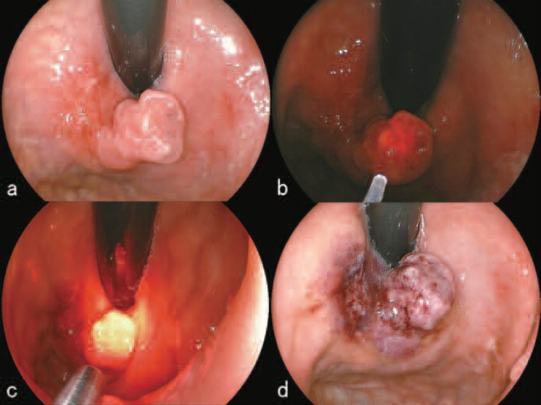
Figure 4:
First treatment course of PDD and PDT using talaporfin sodium (November 10, 2009)
a. Before L-PDD and L-PDT. The anterior portion of the tumor showed a polypoid growth measuring 15 mm in its major axis.
b. L-PDD revealed clear red fluorescence centering on the polypoid tumor.
c. During irradiation with the 664 nm PD laser (Panasonic Healthcare, Tokyo, Japan) using a direct-type quartz fiber.
d. After the irradiation, cessation of blood flow within the tumor was observed and the color of the irradiated area turned dark reddish.

