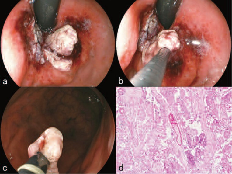
Figure 5:
Three days after the 1st treatment course of L-PDT (November 10, 2009)
a. The size of the polypoid portion of the tumor had decreased and the color of the tumor surface had become whitish.
b. Parts of the tumor were excised with endoscopic snare resection using a high-frequency electric current device.
c. Just after excision.
d. High power magnified view (Hematoxylin-Eosin stain X 100) of the excised tumor. Histopathological examination showed a fibrinoid degeneration of blood vessels surrounded by coagulation necrosis.

