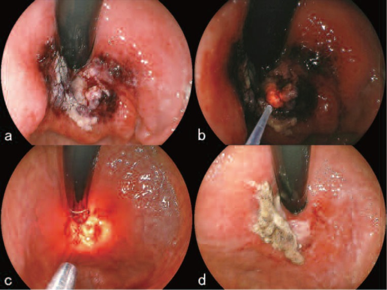
Figure 6:
First course of L-PDD and L-PDT (November 13, 16, 2009)
a. Just after excision. During the excision, virtually no bleeding was seen.
b. L-PDD of the excised area revealed red fluorescence confirming that there was residual talaporfin sodium in the tissue and that cancerous tissue was therefore still there.
c. Additional irradiation with the PD laser using the direct-type quartz fiber.
d. On November 16, endoscopic examination showed a laser ulcer 25 mm in diameter, covered with necrotic debris but without any bleeding.

