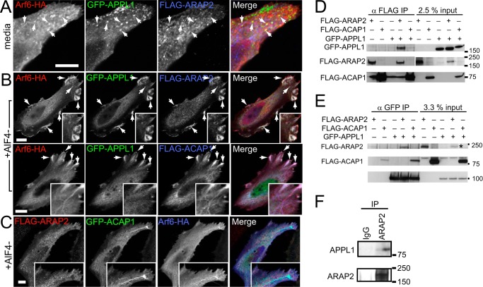
FIGURE 6.
Differential association of ARAP2 and ACAP1 with APPL. A–C, localization of ARAP2 and ACAP1 relative to APPL and to each other. A and B, HeLa cells expressing Arf6-HA, GFP-APPL1, and either FLAG-ARAP2 or FLAG-ACAP1 were treated with (B) or without (A) AlF4− for 30 min, fixed, and stained with anti-HA and anti-FLAG antibodies. C, HeLa cells expressing FLAG-ARAP2, GFP-ACAP1, and Arf6-HA and treated with AlF4− were examined. A higher magnification is shown in insets, and arrows indicate structures where colocalization was detected. Scale bars = 10 μm. D and E, differences in ARAP2-APPL1 and ACAP1-APPL1 complex formation. Lysates from HeLa cells expressing FLAG-ARAP2 or FLAG-ACAP1 alone and in combination with GFP-APPL1 were immunoprecipitated (IP) through the FLAG epitope or GFP followed by immunoblotting using anti-GFP and anti-FLAG antibodies. The asterisk indicates the band representing FLAG-ARAP2 input in double-transfected cells. F, association of endogenous ARAP2 with APPL1. Endogenous ARAP2 was immunoprecipitated from HeLa lysates, followed by anti-APPL immunoblotting.

