
FIGURE 8.
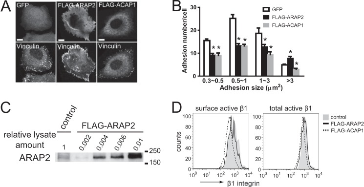
ARAP2 and ACAP1 have different effects on FAs. HeLa cells expressing GFP, FLAG-ARAP2, or FLAG-ACAP1 were plated on fibronectin-coated coverslips for 6 h, fixed, and stained with anti-vinculin and anti-FLAG antibodies. A, representative images. B, adhesion number and size expressed as the mean ± S.E. of at least three experiments. Scale bars = 10 μm. *, p < 0.05. C, levels of recombinant ARAP2 expression relative to endogenous ARAP2. Various amounts of lysates from HeLa cells transfected with FLAG-ARAP2 were analyzed concurrently with 45 μg of lysates from vector-transfected cells by anti-ARAP2 immunoblots to determine fold overexpression of FLAG-ARAP2. D, representative FACS histogram for surface and total active integrin in HeLa cells transfected with vector control, FLAG-tagged ARAP2, or ACAP1.