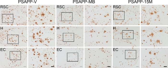
FIGURE 2.

Amelioration of cerebral parenchymal β-amyloid deposits in PSAPP mice treated with methylene blue. Representative photomicrographs were obtained from PSAPP mice treated with vehicle (PSAPP-V) or with MB (PSAPP-MB) for 3 months starting at 15 months of age (mouse age at sacrifice = 18 months) as well as 15-month-old PSAPP mice (PSAPP-15M). Immunohistochemistry using an anti-Aβ17–24 monoclonal antibody (4G8) is depicted, demonstrating cerebral β-amyloid deposits in PSAPP-V, PSAPP-MB, and PSAPP-15M mice. Brain regions shown include the following: RSC (top), H (middle), and EC (bottom). In images from PSAPP-V and PSAPP-MB mice as well as PSAPP-15M mice, each right-hand panel is a higher magnification image from left panel insets. Scale bars, 200 μm (gray) and 100 μm (black).
