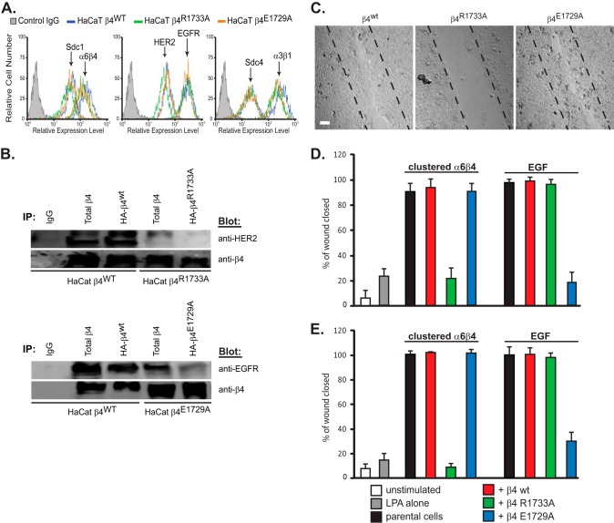
FIGURE 7.
Dominant negative α6β4R1733A integrin disrupts HER2-dependent haptotaxis and α6β4E1729A disrupts EGFR-dependent chemotaxis of HaCat and MCF10A cells. A, HaCat cells expressing β4WT, β4R1733A, or β4E1729A mutants are subjected to flow cytometry to determine relative cell surface expression of α6β4 integrin, Sdc1 and Sdc4, α3β1 integrin, and HER2 or EGFR. B, either total or HA-tagged β4 integrin was immunoprecipitated (IP) with mAb 3E1 or anti-HA from lysates of HaCat cells expressing HA-β4WT, HA-β4R1733A, or HA-β4E1729A mutants. Immunoprecipitates divided into duplicate samples were probed for co-immunoprecipitation of integrin and either HER2 or EGFR. C, confluent HaCat cells expressing either β4WT, β4R1733A, or β4E1729A integrin subunits are stimulated to close a scratch wound during a 15–18-h treatment with 3 μm LPA, 10 μg/ml 3E1, and 30 μg/ml anti-mouse secondary antibody to stimulate HER2-dependent haptotaxis. Bar, 40 μm. D, quantification of scratch wound closure by either parental HaCat cells or cells transfected with β4 integrin constructs and stimulated to undergo haptotaxis or chemotaxis in response to 10 ng/ml EGF. The migration of parental cells in the absence of stimulation or stimulated with LPA alone is shown as a control. E, quantification of scratch wound closure by either parental MCF10A cells or cells transfected with β4 integrin constructs and stimulated as described in C. Data represent the mean of six wells ± S.D.

